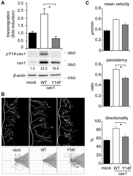
Figure 4. Caveolin-1 aminoacid tyrosine-14 is required for directionality and persistence of migration in B16-F10 cells.
(A) B16-F10 cells transfected with pLacIOP (mock), pLacIOP-caveolin-1 (WT) or the pLacIOP-caveolin-1/Y14F mutant (Y14F) were treated with 1mM IPTG for 24 hours. Then, cell migration was assessed in a Boyden chamber assay by seeding cells (5×10⁴) on fibronectin-coated (2 µg/ml) transwell plates and allowing migration for 2 hours. Cells that migrated to the lower side were detected by crystal violet staining (mean ± SEM, n = 3). *P<0.05. Bottom panels show total protein extracts, separated by SDS-PAGE (35 µg total protein per lane) and analyzed by Western Blotting. Numbers below panels indicate relative caveolin-1 levels (caveolin-1/WT = 22.0±1.8, caveolin-1/Y14F = 16.6±3.8; values compared with the mock condition and obtained as the mean from three independent measurements ± SEM). (B) Confluent monolayers of B16-F10 cells transfected with pLacIOP (mock), pLacIOP-caveolin-1 (WT) or pLacIOP-caveolin-1/Y14F (Y14F) were wounded with a pipet tip and recorded by time-lapse video microscopy for 10 hours (12-min frame interval). Cell tracks were determined as indicated in Figure legend 3B. Tracks of single cells at the wounded edge are shown (upper panel). The edge of the wound is outlined by a white line. Single cell tracks are shown in a Cartesian coordinate system for each cell type (lower panel). (C) The mean velocity ( µm/min) was derived from the instant velocity (Figure S3), as described in (D). The persistency and directionality of migration were obtained as described in Figures 3E and 3F, respectively. Data shown are the mean ± SEM (n = 3). *P<0.05.

