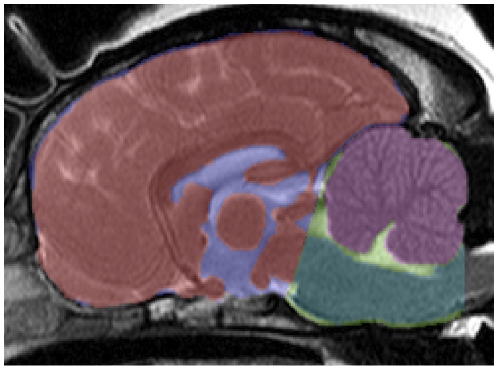
Figure 1. Masks recorded from MR images.
Masks were recorded for the following volumes (mid-saggital view): Parenchyma within the cranial cranial fossa (red), cerebellum (purple), brainstem (dark green), CCF (light green). A mask for the cranial cranial fossa was not recorded but is shown here for completeness (blue).

