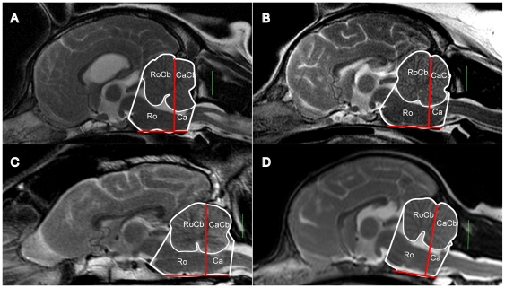
Figure 2. Partitioning of the CCF and cerebellum (mid-saggital view).
A: CKCS - CM/SM group (top left), B: CKCS - CM group (top right), C: Labrador (bottom left), D: small breed dog – Chihuahua (bottom right). Masks were recorded for the following volumes: rostral CCF (Ro), caudal CCF (Ca), rostral cerebellum (RoCb – superimposed on Ro) and caudal cerebellum (CaCb – superimposed on Ca). The green bar is a 1 cm scale. The Red bars represent the division of the CCF into a caudal part and a rostral part by a plane orthogonal to the sagittal images, intersecting the base of the internal occipital protuberance and orientated perpendicular to the basioccipital bone.

