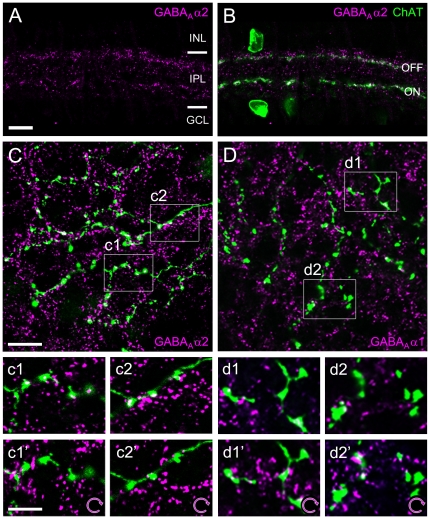
Figure 1. Localization of candidate GABAAR receptor subunits in relation to SAC dendrites and varicosities.
A–B. Immunolabeling pattern of the GABAAR α2 subunit (magenta) in a vertical section of rabbit retina (single optical section; INL, inner nuclear layer; IPL, inner plexiform layer; GCL, ganglion cell layer). Starburst amacrine cells (SACs) were labeled against choline acetyltransferase (ChAT, green). ON SACs have somata in the GCL and their dendrites form the inner ChAT band; OFF SACs are located in the INL and their dendrites form the outer band. Synaptic receptor clusters (magenta) are unevenly distributed across the neuropile, with GABAAR α2 puncta concentrating in two bands along the SAC processes. C. Distal dendrites of a SAC injected with Neurobiotin (green) and co-stained with GABAAR α2 antibody (magenta) (single optical section): the majority of SAC varicosities are associated with receptor staining. Examples for such association are illustrated at higher magnification: c1 and c2 show observed receptor distribution, c1′ and c2′ show randomized control (magenta channel rotated 90° clockwise). Significantly fewer varicosities are associated with receptor clusters in the rotated control. D. Distal dendrites (green) of a SAC injected as in C but co-stained with GABAAR α1 (magenta) (single optical section): Some varicosities are associated with receptor clusters (see also magnifications in d1–d2). No obvious change in the degree of signal overlap is seen for the randomized control (d1′ and d2′). Scale bars: A, 10 µm (applies also to B); C, 10 µm (applies also to D); c1, 5 µm (applies to all insets).

