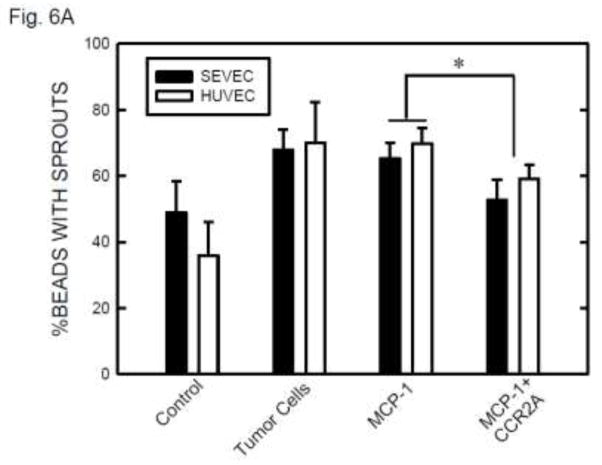
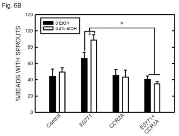
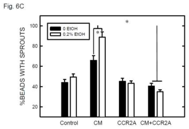
Figure 6.
The role of MCP-1 in ethanol-induced angiogenesis. A: Endothelial cells (SVEC or HUVEC) attached to cytodex beads were suspended in fibrin gel containing MCP-1 (0 or 10 ng) with/without CCR2 antagonist (20 nM). At 12 hours after incubation, the images of endothelial cells with sprouts were captured and the percentage of beads with sprouts was determined as described above. Values were the mean ± SEM of three independent experiments. * denotes a significant difference. B: SVEC cells attached to cytodex beads were suspended in fibrin gel containing CCR2 antagonist (0 or 20 nM). One milliliter of medium containing E0771 cells (2 × 10⁴) and ethanol (0 or 0.2%) was layered on top of the gel. Ethanol exposure was carried out using a sealed container system as described under the Materials and Methods. At 12 hours after incubation, the images of endothelial cells with sprouts were captured and the percentage of beads with sprouts was determined as described above. Values were the mean ± SEM of three independent experiments. * denotes a significant difference. C: E0771 cells were maintained in medium containing 1% FBS and exposed to ethanol (0 or 0.2%) for 24 hours. The conditioned media (CM) were collected and 1 ml of medium was added to fibrin gels containing SVEC cells/beads with/without CCR2 antagonist (CCR2A, 20 nM). At 12 hours after incubation, the images of endothelial cells with sprouts were captured and the percentage of beads with sprouts was determined as described above. Values were the mean ± SEM of three independent experiments. * denotes a significant difference.


