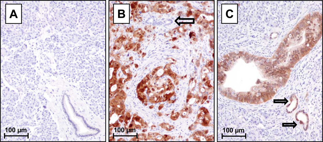
Figure 1.
Immunohistochemical analysis of AKR1B10 expression in human pancreatic tissues: (A) No detectable expression was observed in normal pancreatic acini or ducts. (B) Well-differentiated pancreatic ductal adenocarcinoma with high expression of AKR1B10, but negligible expression in an adjacent normal duct (open black arrow). (C) Precancerous pancreatic epithelial neoplasia also over-expressed AKR1B10 as demonstrated in pancreatic intraepithelial neoplasia-3 (large duct in center) and pancreatic intraepithelial neoplasia-1 lesions (open black arrows).

