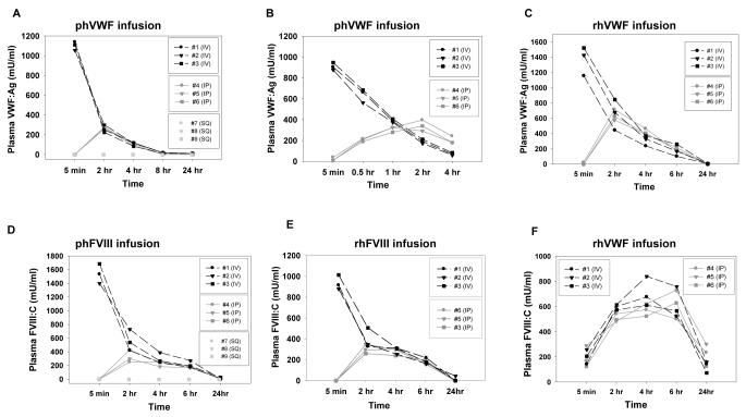
Figure 1. Circulating levels of VWF and FVIII in the infused animals.
Animals were infused with human VWF and /or FVIII from various sources at 50 IU/kg using varying routes. Plasma samples were collected at various time points for VWF:Ag ELISA or FVIII:C assay. Fig. 1A shows the levels of VWF in FVIIInull mice over a 24 hour period after infusion of Alphanate containing plasma derived human VWF (phVWF) and FVIII (phFVIII). A human-specific VWF:Ag ELISA assay was used. Fig. 1B shows the first 4 hours after infusion of Alphanate in another experiment. Fig. 1C shows the levels of VWF in VWFnull mouse plasma after infusion of recombinant human VWF (rhVWF). Fig. 1D shows the levels of FVIII in FVIIInull animals that were infused with Alphanate. Fig. 1E shows the levels of FVIII in FVIIInull mice that were infused with recombinant human FVIII (rhFVIII). Fig. 1 F. shows that endogenous murine FVIII was restored in VWFnull mice when rhVWF was infused whether by IP or IV injection.

