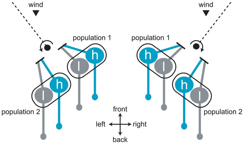
Figure 3. Organization of the wind sensing system in Drosophila.
Wind-sensitive neurons are located in Johnston’s organ, which senses the movement of the most distal antennal segment relative to the rest of the antenna. This figure schematizes one pair of Johnston’s organs as viewed from above the dorsal side of the fly’s ahead. The dashed line represents the plane of the arista, a feather-like structure which protrudes from the distal antennal segment. Wind pushing on the arista rotates the distal antennal segment, and this stretches the dendrites of one population of neurons within Johnston’s organ while compressing the dendrites of the opponent population in the same organ. In this diagram, wind pointing toward the rear of the fly (arrowhead) is rotating the arista as indicated by the arrows, thereby stretching (exciting) population 1 and compressing (inhibiting) population 2. The magnitude of aristal movement should depend on both the velocity and direction of air movement. Each population of Johnston’s organ neurons is thought to contain cells tuned to high-frequency (h) and low-frequency (l) movement.

