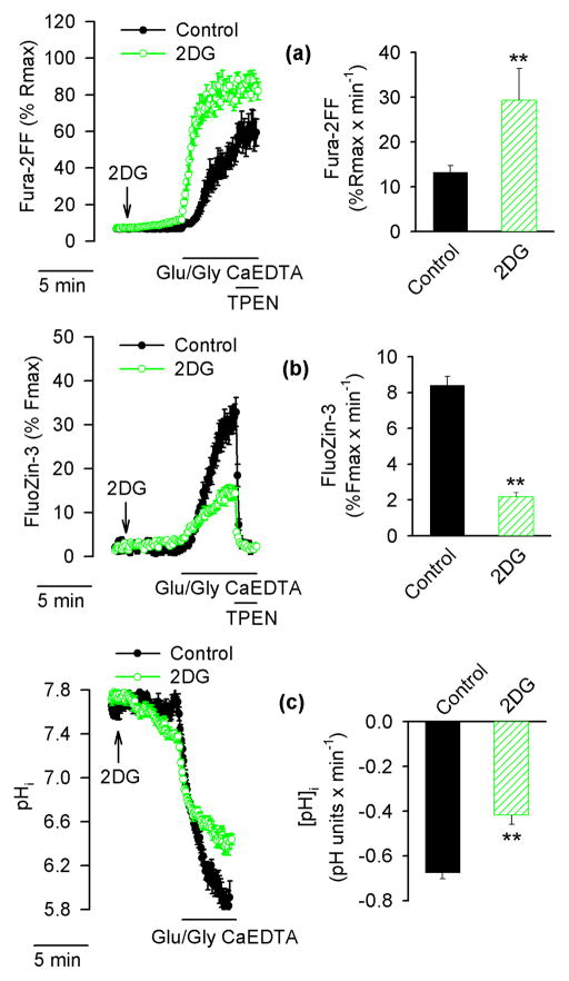
Figure 4.
Effects of substitution of glucose with 2-deoxy-D-glucose (2DG) on [Ca2+]i, [Zn2+]i and pHi in Glu/Gly-treated neurons. Fura-2FF- and FluoZin-3-loaded neurons (a and b) or BCECF-loaded neurons (c) were exposed to Glu/Gly as described in Fig. 1 in the presence of glucose (black traces, filled symbols, black bars) or with glucose substituted with 2DG (green traces, open symbols, hatched bars). The arrow indicates when glucose was replaced by 2DG. The data in the left hand panels are averages ± SE from 13 – 22 neurons monitored in a single experiment; the averages ± SE from 4 such experiments are shown in the right hand panel. ** p < 0.01 (Student’s t-test).

