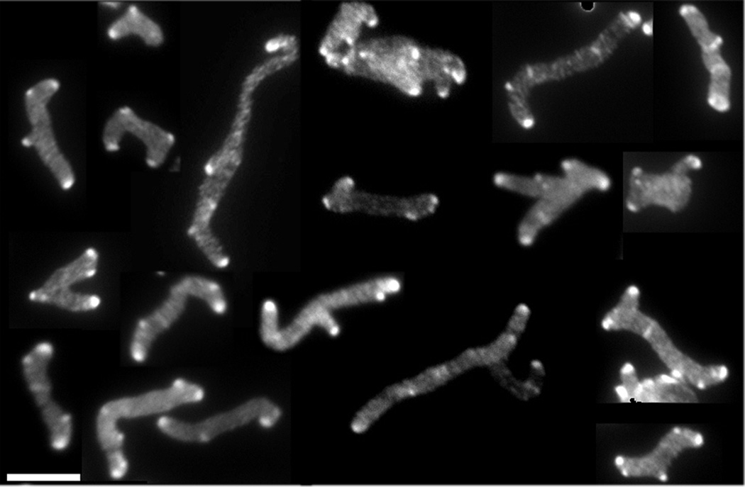
Fig. 3. All branches have inert PG at their poles.
E. coli CS703-1, a mutant lacking PBPs 1a, 4, 5, 6, 7, AmpC and AmpH, was grown in LB containing 100 µg/ml of D-cys for three generations, after which cells were chased in the absence of D-cys by growing them in LB plus 1 µg/ml of aztreonam (to inhibit cell division) for 2 h (~3 mass doublings). After the chase, sacculi were isolated and immunolabelled to detect D-cys residues, as described (de Pedro et al., 1997). This figure is a mosaic of images taken from different fields of view. The scale bar equals 5 µM.

