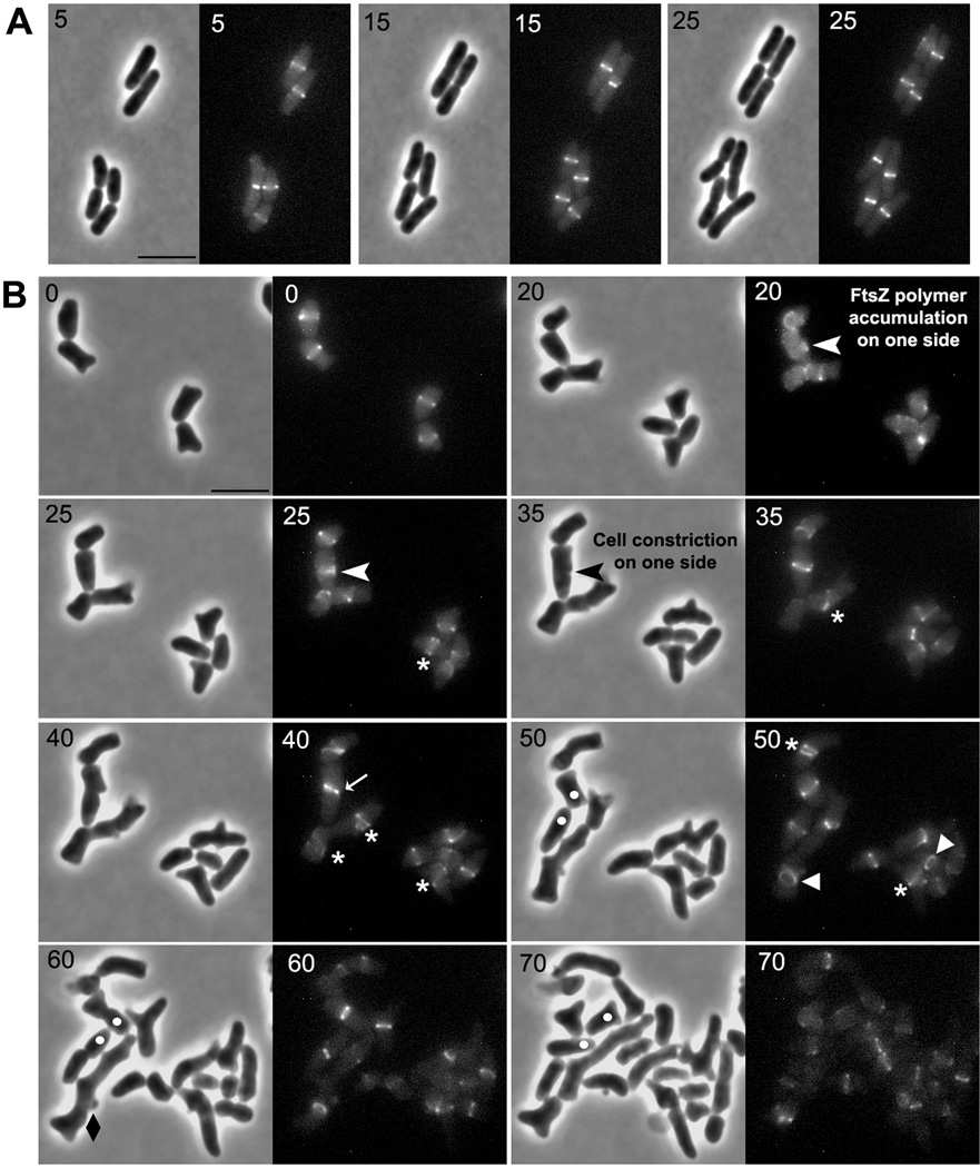
Fig. 6. Abnormal FtsZ polymers are responsible for abnormal cell constriction.
E. coli strains LP18-1 (wild type) (A) and LP1 (Δ PBPs 4, 5 and 7) (B) were grown and imaged as described in Experimental Procedures. White arrow heads in panel B (at time 20 and 25 min) represent abnormal FtsZ polymers that formed on one side of the cell which eventually resulted in abnormal cell constriction (black arrowhead at 35 min). These events gave rise to abnormal poles, leading to the formation of a branch (white dots are inserted to mark and follow the development of selected abnormal cell poles). Abnormal FtsZ polymers are denoted by symbols described in Fig. 5. The numbers in each panel indicate time in minutes. Only selected time points were included in this figure for clarity (see Fig. S6 for a compilation of all time points). The scale bar in panels A and B equals 5 µm.

