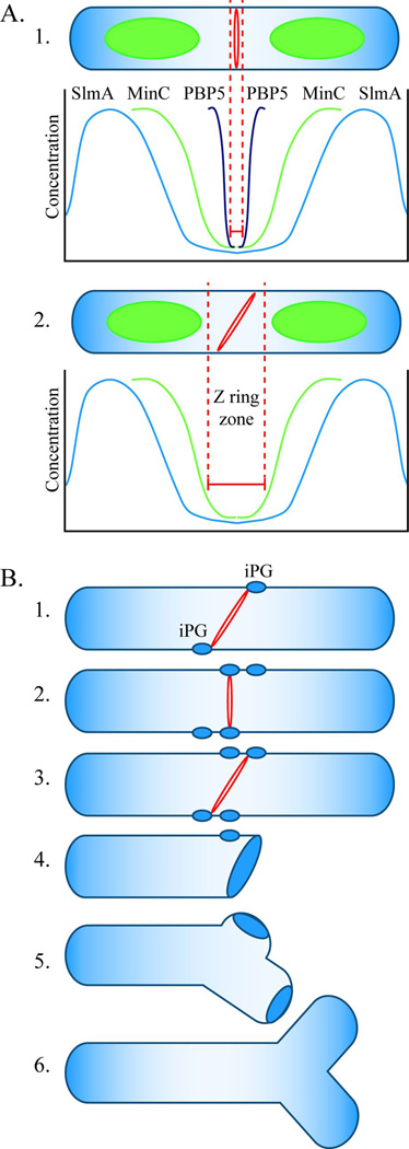
Fig. 9. Model for FtsZ ring localization and cell branching in E. coli.
A. FtsZ ring localization. A1. In wild type E. coli, the FtsZ ring (red ring) is restricted to the center of an elongating cell (“Z ring zone” between dotted red lines) by three mechanisms. The SlmA protein is associated with nucleoids (green ovoids), and its concentration is lowest near the cell’s center and near the poles (blue line). This FtsZ inhibitor restricts Z ring formation to a relatively broad area at these three sites. The MinC protein prevents polymerization of FtsZ at the poles and restricts Z ring formation to a small region near the cell’s midpoint, where the concentration of this inhibitor is lowest (green line). PBP5 (black line) fine tunes the location of Z rings to an even narrower zone at the cell’s center. The action of PBP5 is probably aided by other LMW PBPs. A2. In the absence of PBP5, FtsZ can polymerize in a larger midcell zone to form a slanted Z ring (shown) or multiple Z rings (not shown). Before invagination begins, the orientation of midcell Z rings can vary within the permissive zone. B. Mechanism of cell branching. B1. In the absence of PBP5, slanted FtsZ rings can form at the center of the cell and initiate synthesis of inert peptidoglycan (iPG) (blue ovals). B2. Before invagination begins, the Z ring can reorient and initiate the synthesis of iPG at sites well separated from the first. B3. A non-perpendicular Z ring triggers invagination. B4. The daughter cells have one pole consisting of iPG, plus at least one other patch of iPG some distance away. B5. During subsequent cell growth, insertion of peptidoglycan into the sidewall separates the true pole from the patch of iPG. B6. Cell wall elongation around the iPG patch creates a branch and an ectopic pole.

