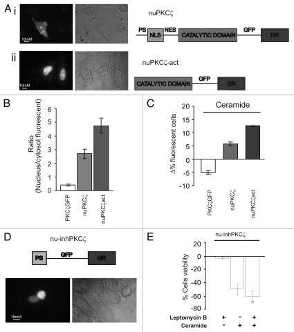
Figure 3.
Effects of constitutively nuclear PKCζ (nuPKCζ), constitutively active and constitutively nuclear PKCζ (nuPKCζ-act) and selectively nuclear PKCζ inhibitor (nu-inhPKCζ) on cell viability. (A) Cellular localization and schematic structure of the chimera PKCζ: (i) nuPKCζ and (ii) nuPKCζ-act. (B) Nuclear localization of the different PKCζ chimeras: the histograms represent the ratio between the nuclear and cytosolic average fluorescence intensity, expressed as a percentage, for the three chimera proteins in resting conditions (nuclear distribution of: nuPKCζ-act 4.71 ± 0.80 or nuPKCζ 2.76 ± 0.41 vs. 0.41 ± 0.03 for PKCζ-GFP). (C) Apoptotic counts of HeLa cells expressing the different PKCζ mutants following ceramide treatment (cell viability in nuPKCζ-act and nuPKCζ cells: Δ% 12.6 ± 0.40 or 6.3 ± 1.08 vs. −5.3 ± 1.11 in PKCζGFP expressing HeLa cells, respectively). (D) Cellular localization and schematic structure of the selectively nuclear PKCζ inhibitor (nu-inhPKCζ). (E) Effects of nu-inhPKCζ overexpression on cell viability following LMB pretreatment and/or C2-ceramide treatment (Δ% cells viability in pre-treated LMB cells ceramide untreated: −4.6 ± 2.31 vs. −60.0 ± 8.60 after ceramide treatment or in not LMB pre-treated −48.6 ± 8.71).

