
Figure 1.
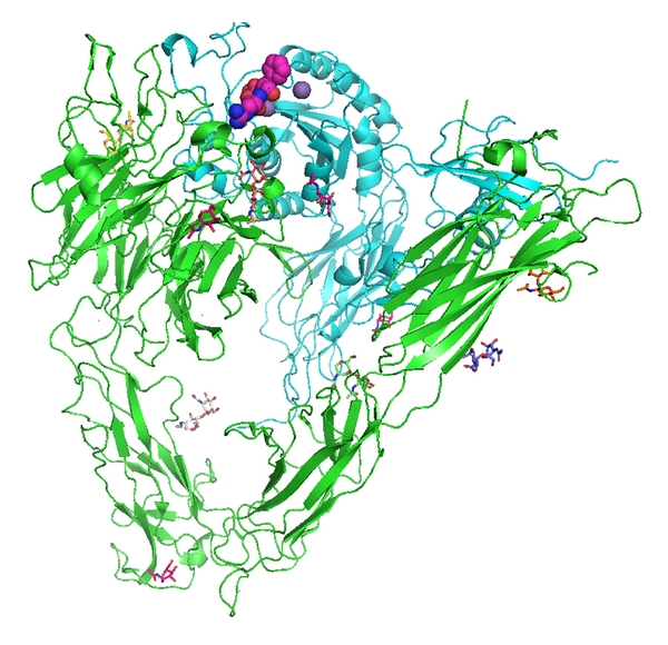
View of αvβ3 integrin from 1L5G.pdb (Xiong et al., 2001 [4]). RGD cyclic pentapeptide is shown as space filling atoms (violet), NAG sugars are stick figures in various colors, and the sphere (purple) is a Mn2+ ion. The αv domain is green and the β3 domain is cyan. Figure drawn with PyMol.