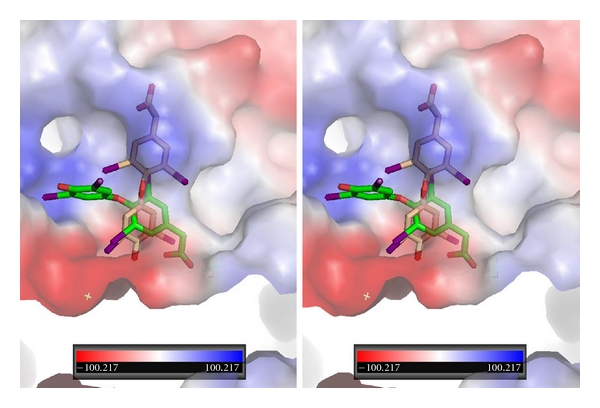
Figure 7.

Stereoview of QM/MM model for T4ac bound in αvβ3 integrin at the RGD binding site. T4ac bound in two orientations: one orientation (green, Ca1) has the phenolic ring binding in a different pocket than that of T4 while the other orientation of T3 (beige, Ca2) binds in a mode that is perpendicular to the first.
