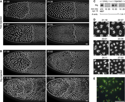
Figure 1.
Cdc25 mRNA injection induces a successful, early mitosis in cycle 14 during what would ordinarily be S phase. (A,B) Stills from movies following His2AvD-GFP embryos injected during cycle 13 with Twe (A) or Stg (B) mRNA. Times displayed are time after mitosis 13. The region undergoing division (dotted line) is localized near the point of injection (left pole). (A) A single mitosis after a 21-min interphase (see Supplemental Movie S1). (B) Two successive induced mitoses—the first (dotted line) after a 23-min interphase, and the second (dashed line in bottom panels) ∼35 min later. The normal onset of gastrulation movements has shifted nuclear positions in the last panel (see Supplemental Movie S2). (C) Western blot showing Stg expression in injected embryos is similar to that in pre-MBT embryos. Lanes were loaded with aliquots equivalent to either one or one-quarter embryo (# emb.) from pooled extracts of three embryos of the indicated type. The pre-MBT sample is from cycle 11 embryos. Time into cycle 14 was estimated by nuclear length. (D) Series of stills from Supplemental Movie S3. These high-magnification views of the induced mitosis in an embryo injected with Stg mRNA show no DNA bridging at anaphase (see especially panels 23:01 and 23:36). Times are those displayed in Supplemental Movie S3. (E) Induced anaphases in a Stg mRNA-injected embryo that was fixed 22 min after mitosis 13. Hoechst staining of the DNA (green) shows no evident bridging of the DNA, and the in situ hybridization signals for the X-chromosomal 359 satellite (red) and the Y-chromosomal AATAC satellite (cyan) show distinct foci of these late-replicating sequences in each segregating complement. Bars, 10 μm.

