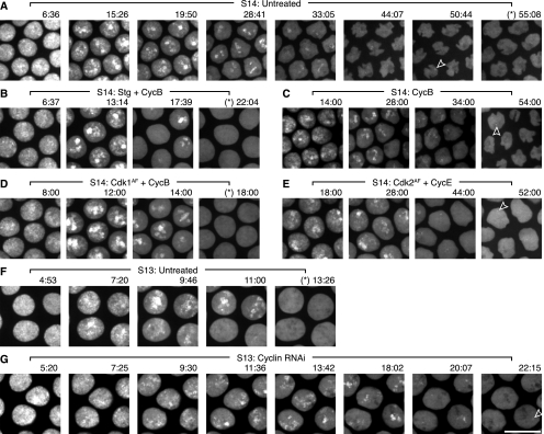
Figure 5.
Stg or Cdk1AF mRNA, coinjected with CycB mRNA, accelerate replication, while dsRNA against mitotic cyclins slows replication. Stills from Supplemental Movie S6. Embryos were filmed after injection with purified GFP-PCNA protein and nothing (A,F), Stg and CycB mRNAs (B), CycB mRNA (C), Cdk1AF and CycB mRNAs (D), Cdk2AF and CycE mRNAs (E), or dsRNA against CycA, CycB, and CycB3 (G). Time into interphase 14 is marked above the micrographs. An asterisk (*) before the time indicates a micrograph in which no PCNA foci are visible. In some panels, an open arrowhead highlights the last visible late PCNA focus. Also, in some later time-point micrographs (e.g., A, 44:07), the nuclei can be observed changing shape, which is a normal consequence of cellularization that is happening during cycle 14. Bar, 10 μm.

