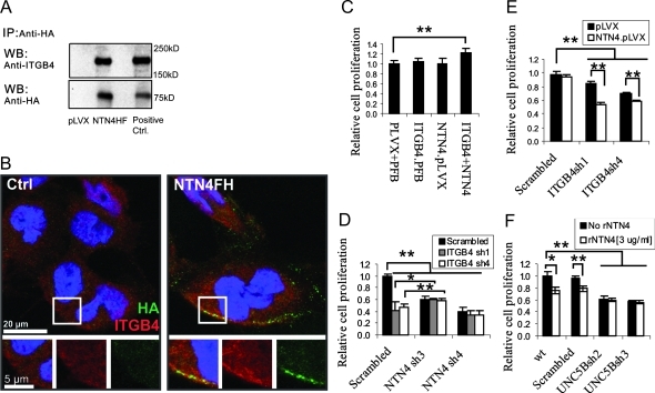
Figure 2.
NTN4/ITGB4 interaction promotes glioblastoma cell proliferation. (A) U251 cells expressing empty vector or FLAG/HA-tagged NTN4 were cultured to 50% confluence and were subsequently lysed for immunoprecipitation. After immunoprecipitation with anti-HA agarose, ITGB4 was detected in immunoprecipitates of FLAG/HA-tagged NTN4. Immunoprecipitates of empty vector and cell lysates were set as negative control and positive control, respectively. (B) U251MG cells were cultured to 50% confluence and were incubated with conditioned medium from either control or FLAG/HA-tagged NTN4 (NTN4FH)-expressing cells as described in Supplemental Materials and Methods, and then used for immunofluorescence. Micrographs of ITGB4 (red) and HA (green) in U251MG were obtained using a confocal laser scanning microscope. Yellow indicates the colocalization of ITGB4 and NTN4. (C) U251 cells expressing the indicated gene or shRNAs were cultured in 96-well plates for 2 days and then used for the proliferation assay. Neither NTN4 overexpression nor ITGB4 overexpression alone promoted U251MG cell growth. Combined overexpression of NTN4 and ITGB4 increased cell proliferation by approximately 30%. (D) U251 cells expressing the indicated gene or shRNAs were used for proliferation assay. Suppression of ITGB4 expression in NTN4-silenced U251MG cells did not further reduce cell proliferation. Complete suppression of NTN4 expression, NTN4sh3, slightly increased the proliferation of ITGB4-silenced U251MG cells. (E) Suppression of ITGB4 expression leads to less mitogenic rate in NTN4-overexpressing U251MG cell than in control cells. (F) Suppression of UNC5B expression in U251MG cells reduced cell mitogenic ability. Exogenous addition (3 µg/ml) of NTN4 inhibited cell proliferation in control cells but not in UNC5B-silenced cells. Mean ± SE, n ≥ 3. **P < .01. *P < .05.

