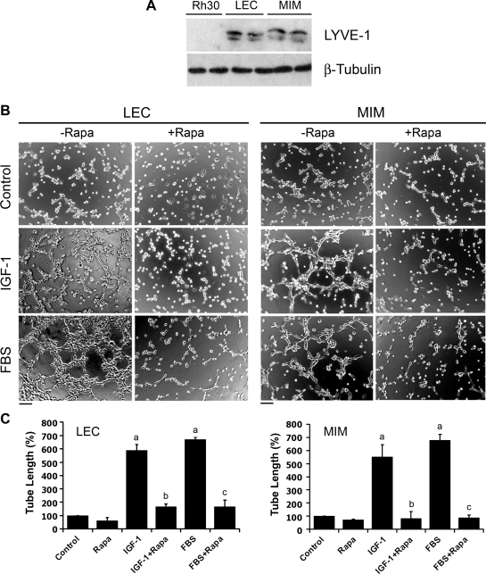
Figure 1.
Rapamycin inhibits IGF-1/FBS-stimulated tube formation in LECs. (A) Cell lysates from indicated cells were subjected to Western blot analysis with the indicated antibodies. (B and C) LEC and MIM cells were treated with rapamycin (Rapa, 100 ng/ml) for 24 hours, in the presence or absence of IGF-1 (10 ng/ml) or 2% FBS, followed by tube formation assay. Representative images are shown in B. Bar, 100 µm. The length of tube-like formation was evaluated by ImageJ software. Quantitative data are presented as mean ± SD (n = 3) in C. aP <.05, difference versus control group. bP < .05, difference versus IGF-1 group. cP < .05, difference versus FBS group.

