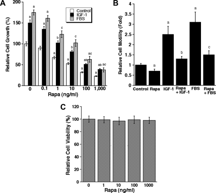
Figure 2.
Rapamycin inhibits proliferation and motility in LECs. (A) LECs, grown in serum-free DMEM/F12 or the medium supplemented with IGF-1 (10 ng/ml) or FBS (10%), were exposed to rapamycin (0-1000 ng/ml) for 72 hours, followed by cell counting using a Beckman Coulter counter. (B) Cell motility of LECs was determined using the single-cell motility assay. (C) LEC viability was evaluated by one-solution assay. Quantitative data are presented as mean ± SD (n = 3) in A to C. aP < .05, difference versus control group. bP < .05, difference versus IGF-1 group. cP < .05, difference versus FBS group.

