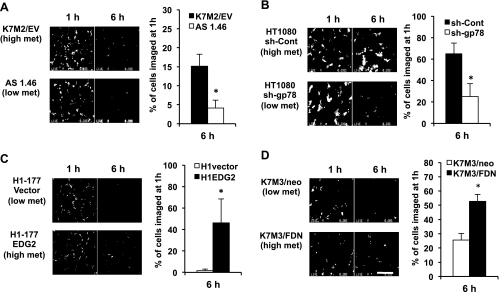
Figure 1.
SCVM reliably distinguished high versus low metastatic (breast cancer and sarcoma) cells within 6 hours of their arrival in the lung. SCVM allows in vivo or ex vivo imaging of single fluorescently labeled metastatic cells in the mouse lung [13]. SCVM findings were seen in a variety of cancer types and biologic settings. (A) Ezrin in murine osteosarcoma (*P = .0043) [13]. (B) E-3 ligase in soft tissue sarcoma (*P = .0004) [20]. (C) EDG2 in human breast cancer (*P = .0056) [21]. (D) FAS-FAS-ligand in murine osteosarcoma (*P = .0007) [22]. Scale bars, 400 µm. Of the high and low metastatic cell line pairs examined, only the HOS-MNNG/HOS pair could not be distinguished at this early time point (Figure W1). All data represent the mean values ± SD from experiments performed at least three times; nonparametric two-tailed t test.

