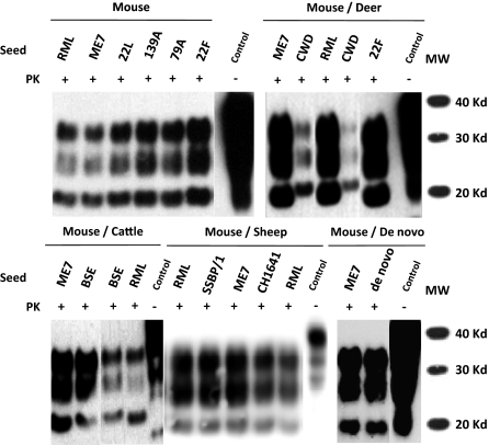
Fig. 1.
Biochemical analysis of different rabbit PrPres (RaPrPres) generated in vitro by serial automated PMCA (saPMCA) using a New Zealand White rabbit brain homogenate as substrate. Rabbit brain homogenates seeded with different prion strains (mouse: RML, ME7, 22L, 139A, 79A and 22F, mule deer: CWD, cattle: BSE, sheep: SSPB/1 and CH1641) or unseeded (de novo) were subjected to saPMCA. Seeded samples from round 10 and the unseeded sample from round 20 were digested with 100 μg/mL of proteinase K (PK) and analyzed by Western blot using monoclonal antibody D18. Three differential electrophoretic migration patterns are shown, depending on the seed that was used: (i) the pattern for the unglycosylated band that migrated the farthest (18–19 Kd, sheep and mouse adapted scrapie strains); (ii) the intermediate migration pattern for the same band (19–20 Kd, BSE); and (iii) the migration pattern for the unglycosylated band that migrated the least (20-21 Kd, CWD). RML and ME7 RaPrPres samples are used in all blots as reference for determining the different electrophoretic migration patterns. Control, normal rabbit brain homogenate.

