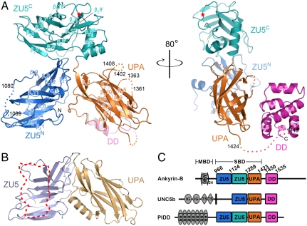
Fig. 1.
Overall structure of ankyrin-B ZZUD. (A) Ribbons diagram representation of the crystal structure of ankyrin-B ZZUD with ZU5N (blue), ZU5C (cyan), UPA (orange), and DD (purple) drawn in their specific colors. The same color code is used throughout the rest of the figures. The disordered regions are indicated by dotted lines with starting and end residues number labeled. The nine-residue deletion site is indicated by a red star. (B) Comparing with ankyrin-B, the ZU5 and UPA domains of UNC5b interact with each other following a similar manner. The DD binding region in ZU5 of UNC5b is highlighted with a dotted trace. (C) Domain architectures of the ZU5-UPA-DD containing proteins in the eukaryotes genomes. The domain boundaries of ankyrin-B ZZUD are defined based on the structure determined in this work.

