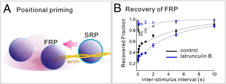
Fig. P1.
(A) Conceptual diagram for positional priming. In this process, reluctant vesicles belonging to the slow-releasing pool (SRP) are converted into fast-releasing vesicles. Synaptic vesicles in the fast-releasing pool (FRP) reside within the Ca2+-microdomain (red hemisphere), whereas vesicles at the periphery belong to the slow-releasing pool. Positional priming is illustrated as an actin-dependent movement of vesicles toward the Ca2+ source. (B) Time courses for changes in sizes of the fast-releasing pool and the slow-releasing pool after specific depletion of the fast-releasing pool. Note that the recovery of the rapid fast-releasing pool (closed circles) is paralleled by a concomitant dip of the slow-releasing pool (open circles) under control conditions. Actin disruption (blue circles) abolished both the rapid recovery phase and the dip in the slow-releasing pool.

