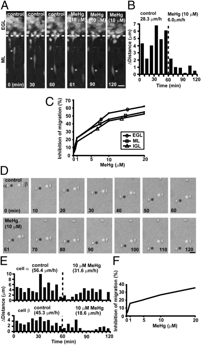
Fig. 2.
MeHg slows granule cell migration in vitro. (A) Time-lapse images showing that 10 μM MeHg inhibits the radial migration of a granule cell in the ML of a cerebellar slice obtained from a P10 mouse. Asterisks mark the granule cell soma. Elapsed time is indicated on the bottom of each photograph. (Scale bar: 12 μm.) (B) Sequential changes in the distance traveled during each 10-min interval by the granule cell shown in A were plotted as a function of elapsed time before and after the application of 10 μM MeHg. (C) Inhibition of the speed of granule cell migration in the EGL, ML, and IGL by MeHg (0.1–20.0 μM). (D) Time-lapse images showing that 10 μM MeHg slows the migration of isolated granule cells in the microexplant cultures of P3 mouse cerebella. Black and white asterisks mark the somata of migrating granule cells. Elapsed time is indicated on the bottom of each photograph. (Scale bar: 12 μm.) (E) Sequential changes in the distance traveled by two granule cells (α and β shown in D) during each 5-min interval were plotted as a function of elapsed time before and after the application of 10 μM MeHg. (F) Inhibition of the speed of migration in the microexplant cultures by MeHg (0.1–20.0 μM).

