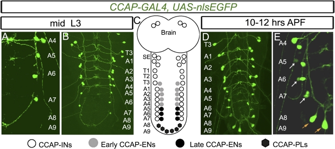
Fig. 1.
Emergence of late CCAP neurons in the A5–A9 abdominal VNC at pupariation. Expression of CCAP-GAL4,UAS-nlsEGFP (green) in the T3–A9 hemisegments in mid-L3 larvae (A and B) and in pupae 10–12 h APF (D and E) is shown with a summary depicting all CCAP neuronal subsets (C). Within each segment (T1–A7), CCAP neurons project across the midline forming a ladder-like structure that we used to confirm the segment identity of every CCAP neuron (see also Fig. S1). (A and B) At mid-L3, there is a CCAP neuron doublet in the T3–A4 hemisegments. In A5–A7 hemisegments, there is only a single CCAP neuron. (D and E) By 10–12 h APF, a second CCAP neuron has emerged in each A5–A7 hemisegment (white arrow). Also, six CCAP neurons emerge within hemisegments A8 and A9 (orange arrow). (C) Cartoon summary of CCAP neurons in the CNS. Based on previous work (25) and the identification of late CCAP-ENs and CCAP-PLs in Fig. 2, we summarize the identity of each CCAP neuron subtype here. Genotype: CCAP-GAL4,UAS-nlsEGFP/+.

