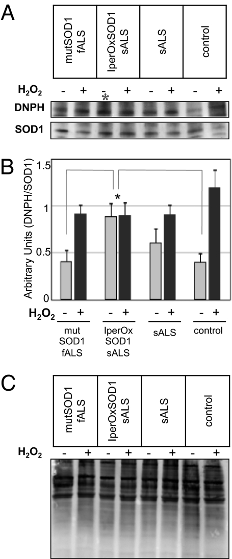
Fig. 1.
Identification of an iper-oxidized SOD1 in a subset of sALS patients. (A) Representative immunoblot of SOD1 oxidation detected by a derivatization assay of carbonyl compounds. Proteins extracted from sALS, fALS, and healthy control lymphoblasts, before and after H2O2 treatment, were precipitated with rabbit anti-SOD1 antibody and analyzed by Western blot using anti-DNPH (Upper) and sheep anti-SOD1 (Lower) antibodies. (B) Ratio of DNPH and precipitated SOD1. For each patient's line, samples were analyzed in triplicate in three independent blots (with and without H2O2), and densitometric analysis was performed on each blot. The graph represents the mean ± SD SOD1 oxidation for each experimental group. ANOVA revealed significantly higher oxidized SOD1 in a group of sALS lymphoblasts compared with lymphoblasts from mutSOD1-fALS and healthy controls (*P < 0.05). (C) Representative blot of derivatization assay of carbonyl compounds on total protein content from fALS, sALS, and healthy control lymphoblasts before and after H2O2 treatment, showing no differences in total oxidation levels.

