Fig. 3.
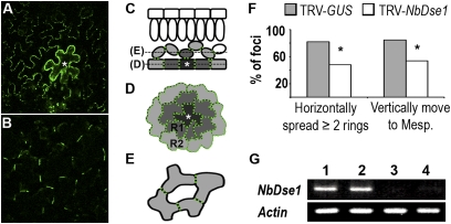
Intercellular movement of P30-1xGFP in DSE1 silenced N. benthamiana leaves. NbDSE1 was silenced by infiltration with TRV-NbDSE1. Plants infiltrated with TRV-GUS were used as controls. (A–E) P30-GFP movement from a primary transformed epidermal cell in N. benthamiana leaves. (A) Leaf epidermis showing horizontal movement from a primary transformed epidermal cell (marked with white asterisk). (B) P30-GFP also moves vertically from the same primary transformed cell into the mesophyll layer of cells. (C–E) Illustration of horizontal and vertical movement of P30-GFP in leaf tissue. (C) Side view of leaf tissue. (D) Top-down view of epidermal cells. (E) Top-down view of spongy mesophyll cells. P30-GFP moves from primary transformed epidermal cells (dark gray with star) to neighboring cells (light gray) and forms puncta (green dots) at PD in the cell wall of the cells to which it moves. In D, R1 and R2 represent the first and second rings of epidermal cells surrounding primary transformed foci. (F) Both horizontal and vertical movements of P30-GFP were reduced in NbDSE1-silenced leaves. Horizontal movement was recorded as the number of rings of epidermal cells into which P30-GFP spread from primary foci. Vertical movement was recorded when P30-GFP moved to mesophyll cells. *P < 0.05 compared with controls. (G) Semiquantitative RT-PCR. NbDSE1 and Actin were amplified in 32 and 25 cycles, respectively. Lanes 1 and 2 are from control TRV-GUS–infected leaves; lanes 3 and 4 are from NbDSE1-silenced leaves.

