
Figure 4.
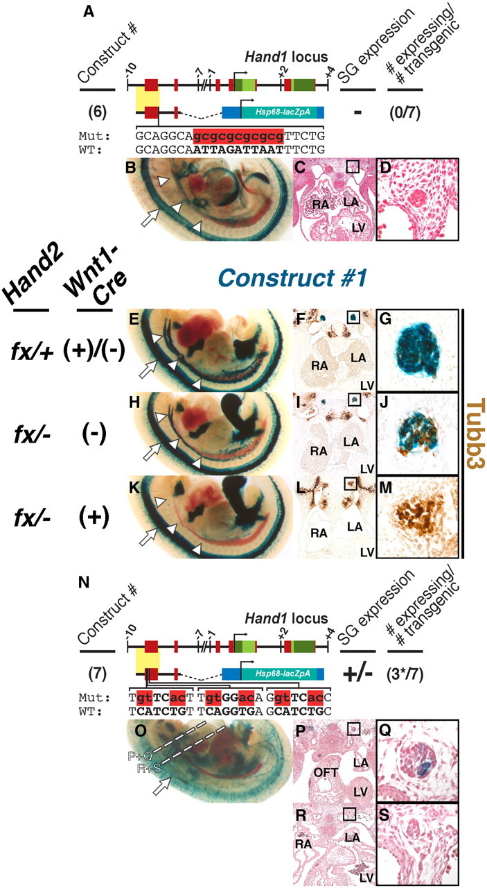
Phox2 and Hand2 are necessary for Hand1-reporter trans-activation in vivo. A, Schematic of Hand1-reporter Construct #6, in which the Phox2 binding site in Construct #4 has been mutated (red). B–D, Zero of 7 E11.5 transient transgenic embryos displayed SG expression. E–M, Expression of Construct #1 is reduced in E11.5 Hand2fx/−;Wnt1-Cre(−) hypomorphs and is lost or reduced in identically staged Hand2fx/−;Wnt1-Cre(+) conditional knock-outs. Cleared whole-mount preparations (E, H, K) show that, compared with control embryos (E), fewer cells within the Hand2 hypomorphic SG (H) express Construct #1 (n = 9 of 10). In the majority of Hand2 conditional knock-out embryos examined, Construct #1 expression is undetectable (K; n = 4 of 6). Transverse sections of Construct #1-expressing embryos, stained with X-gal (blue) in which neurons have been labeled with α-Tubb3 immunohistochemistry (brown), photographed at low (F, I, and L) and high (G, B, and K; denoted by boxes) magnifications show that, whereas SG in control embryos (F, G) are almost exclusively comprised of Hand1-reporter-positive neurons, and Hand2 conditional knock-out SG (L, M) are exclusively comprised of Hand1-reporter-negative neurons, the Hand2 hypomorphic SG (I, J) are comprised of a mix of both Hand1-reporter-positive and -negative neurons. n = 2 for each genotype. N, Schematic of Hand1-reporter Construct #7, in which the three evolutionarily conserved E-box binding sites in Construct #4 have all been mutated (red). O–S, Three of 7 E11.5 transient transgenic embryos displayed detectable SG expression. However, expression in these three embryos was not uniform, and was restricted to SG rostral to the OFT (P, Q). Expression was absent in SG caudal to the OFT (R, S). Dashed lines denote the planes of section for P–S. LA, Left atrium; LV, left ventricle; RA, right atrium.