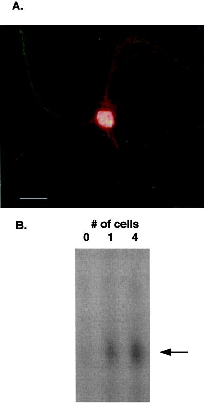
Figure 5.
(A) Rat hippocampal neurons in primary cell culture (36) were fixed in 4% paraformaldehyde in 100 mM Na cacodylate or PBS with 5 mM MgCl2 for 15 min at room temperature and permeablized in 0.1% Triton X-100 in PBS for 15 min at room temperature. Nonspecific interactions were blocked by incubation in 10% normal goat serum, 0.5% nonfat dried milk, 0.1% gelatin, 0.1% Tween-20 in PBS for 2 h at room temperature. Primary Ab was added at 1:150 dilution in blocking solution and incubated overnight at 4°C. Cy3-conjugated secondary Ab was used to visualize 7.16.4 staining (red). Dendritic processes are immuno-stained with MAP2 (green) whereas nuclei are counterstained with 4′,6-diamidino-2-phenylindole (white). (B) These are the IDAT results for lysates isolated from one cell or pooled from four individual cells. The IDAT procedure was as described; 0 cell means that media alone were aspirated into the pipette.

