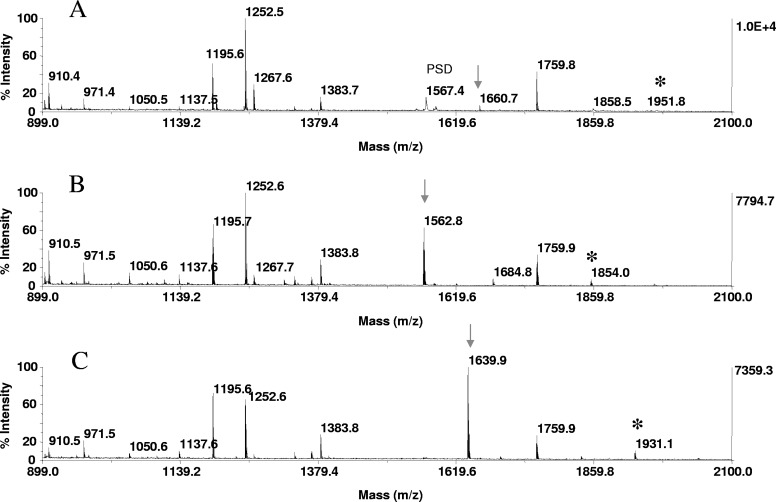
FIGURE 5.
Phosphopeptide detection by signal enhancement. In-gel tryptic digests of α-S1 casein, derived from 20 pmoles protein, loaded onto the gels, were bound to ZipTipC18 pipette tips and subjected to on-resin β-elimination and β-elimination with concurrent Michael addition under the conditions described in Fig. 1. One-tenth of the eluates corresponding to ∼2 pmoles digest was applied to the target. MALDI-TOF spectra of (A) untreated digest, (B) after β-elimination, and (C) after β-elimination with concurrent Michael addition. (A–C) Arrows indicate the phosphoserine peptide at m/z 1660.7, residues 121–134 (VPQLEIVPNpSAEER), its dehydroalanyl derivative at m/z 1562.8, and its thiol adduct at m/z 1639.9, which are observed at an estimated 11- and ninefold higher signal intensity, respectively, relative to the native counterpart. (A–C) *A miscleavage product at m/z 1951.8 spanning residues 119–134, its dehydroalanyl, and thiol derivative.

