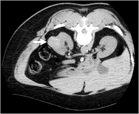
Figure 1.
A 65-year-old woman with left-sided renal mass. Axial noncontrast computed tomography (CT) image performed prior to percutaneous ablation reveals 3.6-cm-diameter high attenuation exophytic mass (arrow) arising from posterior lateral aspect of left kidney. CT-guided biopsy confirmed papillary renal cell carcinoma.

