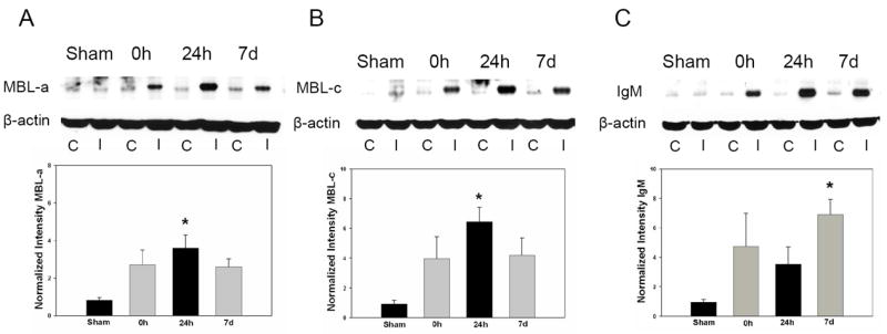
Figure 1. Cerebral ischemia/reperfusion results in a rapid induction of MBL-a, MBL-c, and IgM antigen deposition in the ischemic hemisphere.

Representative Western blots for MBL-a, MBL-c, and IgM were performed in wild-type mice subjected subjected to sham surgery and following 0h, 24h, or 7 days of reperfusion. C= Contralateral hemisphere, I= Ipsilateral hemisphere. Densitometric analyses represent a composite of multiple independent replicates(n=4-5/time-point). (A) MBL-a protein expression increases acutely in the ischemic hemisphere following cerebral ischemia/reperfusion, with a >3-fold increase in MBL-a at 0h relative to sham animals and a peak at 24h(*p<0.05 vs. sham). (B) MBL-c demonstrates a similar(>6-fold) increase by the point of reperfusion, with a similar peak following 24 hours(*p<0.05 vs. sham). (C) Deposition of IgM was also observed to occur rapidly along a similar time-course, but with a peak at 7 days(*p<0.05 vs. sham).
