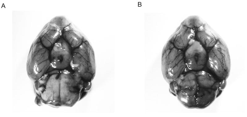
Figure 6. Comparison of cerebrovascular anatomy in mice deficient in MBL-a/c and wild-type controls.

Methylene blue staining of the cerebrovascular anatomy from a ventral perspective reveals no gross anatomic differences in the vascular pattern of the cerebral circulation between (A) MBL-KO and (B) WT mice (representative photograph of n=3 per cohort). Importantly, both genotypes demonstrate bilaterally patent posterior communicating arteries.
