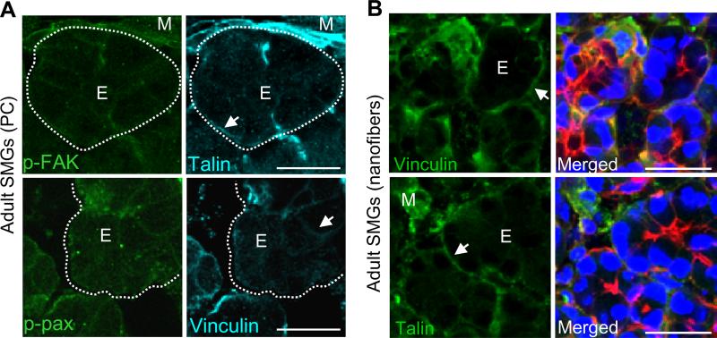
Fig. 2. FA formation and expression is low in adult SMG acinar epithelial cells.
(A) Confocal images of adult SMG tissue explants grown on polycarbonate (PC) for 48hrs ex vivo show low expression of the FA proteins p-FAK, talin, p-paxillin or vinculin within acinar epithelial cells (E, arrows). Staining is seen at the basal cell membranes (arrows) and in mesenchymal cells (M) surrounding the acini, which is outlined by dotted lines. (B) Confocal images of adult SMG tissue explants cultured for 48hrs ex vivo on nanofibers and stained for talin or vinculin (green) or co-stained with actin (red) and DAPI (nuclei, blue) also show low FA expression by acinar epithelial cells with stronger staining at the basal cell membranes (arrows) and in mesenchymal cells. Scale bars = 25 μm.

