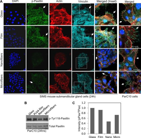
Fig. 4. Phosphorylated paxillin and total vinculin expression are reduced in salivary gland epithelial cells grown on nanofiber scaffolds.
(A) Confocal images of SIMS or ParC10 cells stained for nuclei (DAPI, blue), phospho-Tyr118-paxillin (p-paxillin) (green), actin (red) and vinculin (cyan) indicate FA complex aggregation at cell protrusions (arrows) on the flat or microfiber surfaces which are reduced on nanofiber surfaces. (B) Western blots and (C) densitometric quantification for p-paxillin normalized to total paxillin.

