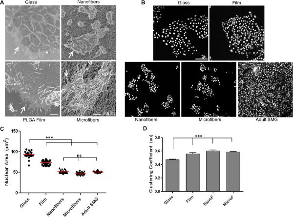
Fig. 5. Nanofiber scaffold topography directs epithelial cell morphology and clustering.
(A) Scanning electron micrographs show cell attachment, protrusions (arrows), and spreading of SIMS cells on glass, spin-coated PLGA film, nanofibers or microfibers, scale bars =20 μm. (B) Binarized segmented confocal images of DAPI-stained nuclei from SIMS cells on different surfaces or adult SMG tissue thin sections, scale bar = 100 μm. (C) Quantification of nuclear area as an indirect measure of cell spreading shows statistically significant differences between cells on flat versus fiber surfaces. No significant difference (ns) was detected between cells on fibers versus adult tissue, *p<0.05 was considered statistically significant using ANOVA followed by Bonferroni's post hoc tests to compare individual means. (D) Quantification of cell clustering using cell-graphs shows an increasing trend in cell clustering (au= arbitrary units) on fiber surfaces compared to glass (***p<0.0001, ANOVA).

