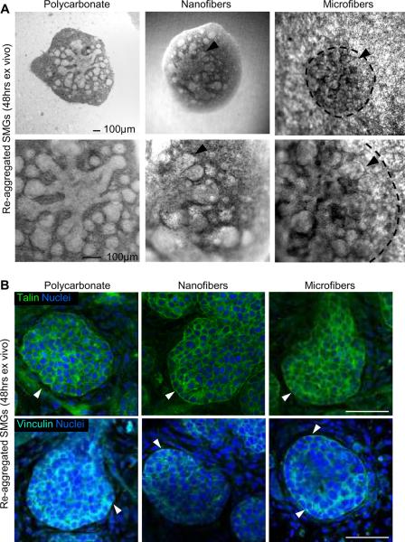
Fig. 6. Nanofiber scaffolds promote self-organization and branching of dissociated embryonic salivary gland cells.
(A) Brightfield images of spontaneously re-aggregated cell pellets from dissociated E13 mouse SMGs cultured ex vivo for 48 hrs on PLGA fibers show extensive bud outgrowth (arrowheads) and ductal elongation, similar to those cultured on a polycarbonate membrane, scale bars = 100 μm. The morphology of re-aggregated structures on microfibers is outlined by dashed lines to compensate for the interference with light microscopy. (B) Confocal images through the equatorial section of re-aggregated buds at 48 hrs stained for talin (green, top panels) or vinculin (cyan, bottom panels) and co-stained for nuclei (DAPI, blue) show diffuse cortical expression with stronger staining along the basal cell membranes at the bud periphery (arrowheads), similar to that observed in intact glands, scale bars = 50 μm.

