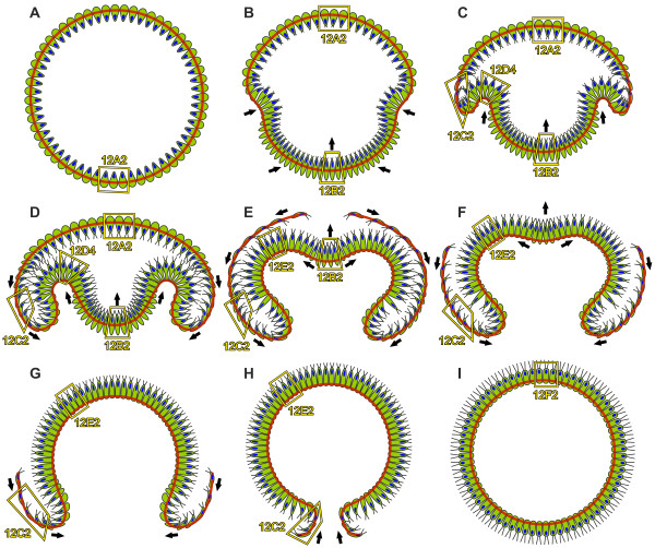
Figure 13.
Model of the inversion process in V. globator. A diagrammatic representation of inversion as deduced from the LM, SEM, TEM and cLSM analyses showing 2D midsagittal cross sections of inverting embryos. Cell content is shown in green; red lines indicate the position of the CB system; and nuclei are shown in blue. Frames show examples of the location of the cell shapes represented in 3D in Figure 12. The directions of the cell layer movements are indicated by black arrows. (A) Pre-inversion stage. (B, C) Early inversion stage; inversion begins with the contraction of the posterior hemisphere and the formation of the bend region. (D to F) Mid-inversion stage; the posterior hemisphere continuously moves into the anterior hemisphere and inverts at the same time; the phialopore opens at the anterior pole and widens; and the anterior hemisphere begins to move over the already inverted posterior hemisphere and inverts at the same time. (G, H) Late inversion stage; the anterior hemisphere continues to move over the already inverted cell monolayer; and inversion ends with the closure of the phialopore. (I) Early post-inversion stage. CB: cytoplasmic bridge; cLSM: confocal laser scanning microscopy; LM: light microscopy; SEM: scanning electron microscopy; TEM: transmission electron microscopy.

