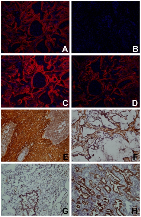
Figure 7. Thrombospondin-2 (TSP-2) histochemical identification in tissue samples.
TSP-2 is identified in serial frozen sections of a single lung carcinoma specimen by (A) a home-made rabbit polyclonal TSP-2 polyclonal antibody, (B) the pre-immune serum from rabbits used to make the home-made polyclonal antibody, (C) a commercial (Novus) rabbit polyclonal TSP-2 antibody, and (D) the TSP-2 SOMAmer. The TSP-2 SOMAmer was used to stain frozen sections of normal and malignant lung tissue, with standard Avidin-Biotin-Peroxidase color development, to demonstrate different morphologic distributions: (E) Strong staining of the fibrotic stroma surrounding tumor nests, with minimal cytosolic staining of carcinoma cells, (F) Strong staining of the fibrotic stroma surrounding tumor nests in a mucinous adenocarcinoma, with no significant staining of the carcinoma cells, (G) normal lung tissue, showing strong cytosolic staining of bronchial epithelium and scattered alveolar macrophages, and (H) strong cytosolic staining of an adenocarcinoma, with no significant staining of the non-fibrotic, predominantly inflammatory stroma.

