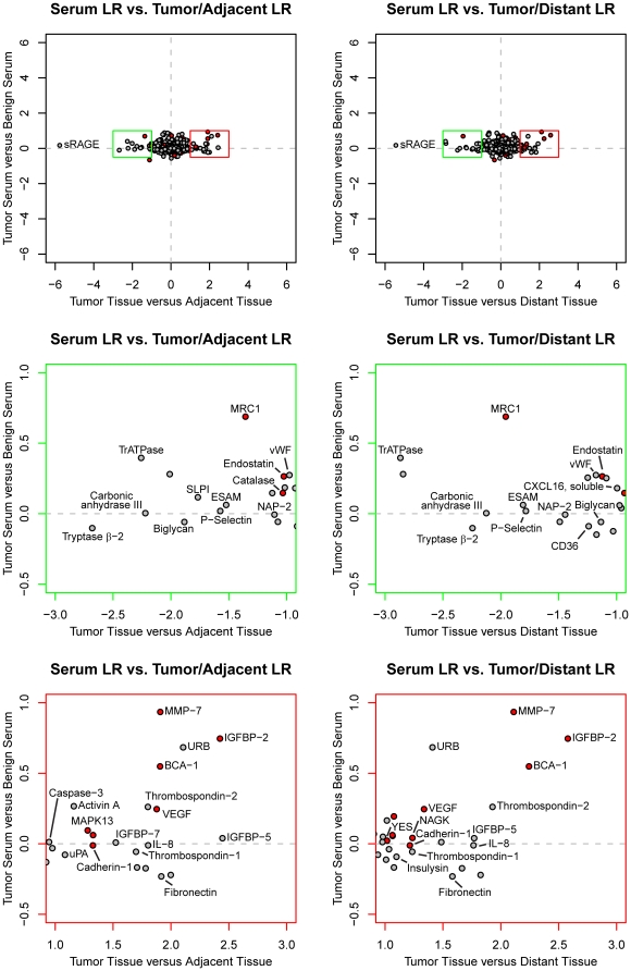
Figure 9. Changes in protein expression in NSCLC tissue compared to serum.
The top two panels show the log2 ratio (LR) derived from serum samples versus log ratios derived from adjacent tissue and distant tissue, respectively. The bottom four panels feature zoomed portions of plots above, indicated by the color of the plot (green for decreased and red for increased expression compared to non-tumor tissue). Analytes shown in Figure 2 have been labeled and analytes mentioned in the publication on the serum samples are shown in filled red symbols red.

