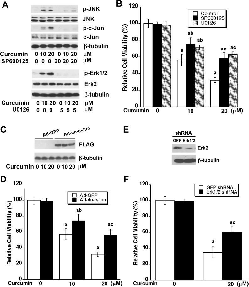
Fig. 3.
Inhibition of JNK or Erk1/2 attenuates curcumin-induced cell death. (A and B) Inhibition of JNK or Erk1/2 with selective inhibitors attenuates curcumin-induced cell death. Rh30 cells were pretreated with SP600125 (20 μM) or U0126 (5 μM) for 30 min and then exposed to curcumin (0–20 μM) for 24 h (for western blotting) or 48 h (for cell viability assay). (A) Western blot analysis was performed with indicated antibodies. (B) Cell viability was evaluated using one solution reagent. Results are presented as mean ± SE (n = 6). aP < 0.05, difference versus control group; bP < 0.05, difference versus 10 μM curcumin group; cP < 0.05, difference versus 20 μM curcumin group. (C–F) Overexpression of dominant negative c-Jun or downregulation of Erk1/2 attenuates curcumin-induced cell death. Rh30 cells, infected with recombinant adenovirus encoding FLAG-tagged dominant negative c-Jun (Ad-dn-c-Jun) or GFP (control) for 24 h, were exposed to curcumin (0–20 μM) for 24 h (for western blotting) or 48 h (for cell viability assay). (C) Western blot analysis was performed with indicated antibodies. (D) Cell viability was evaluated using one solution reagent. Results are presented as mean ± SE (n = 6). aP < 0.05, difference versus control group; bP < 0.05, difference versus 10 μM curcumin group with Ad-GFP infection; cP < 0.05, difference versus 20 μM curcumin group with Ad-GFP infection. Rh30 cells were infected with lentiviral shRNA to Erk1/2 or GFP for 5 days, followed by western blotting with indicated antibodies (E) or further exposed to curcumin (0–20 μM) for 48 h, followed by cell viability assay using one solution reagent (F). Results are presented as mean ± SE (n = 6). aP < 0.05, difference versus control group; bP < 0.05, difference with 20 μM curcumin group with shRNA-GFP infection.

