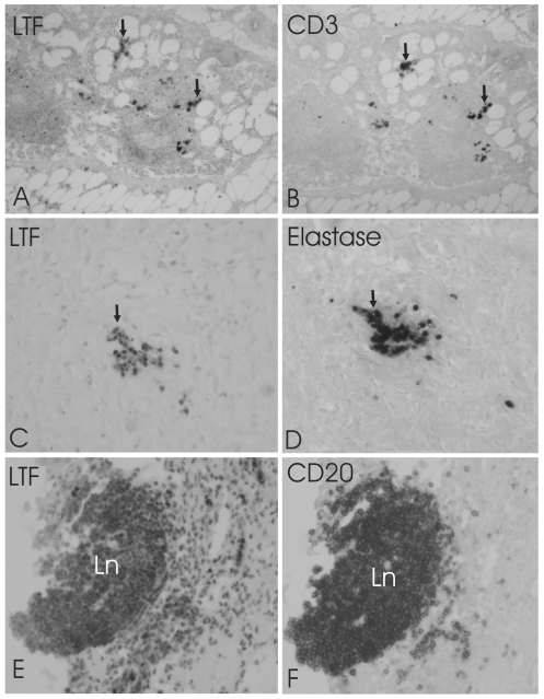
Figure 2. Co-localization of lactoferrin (LTF) with neutrophils and T and B lymphocytes.
Adjacent mirror image sections demonstration the co-localization of LTF (A, C, E) with T-cell marker CD3 (B), neutrophil granulocyte marker elastase (D) and B-cell marker CD20 (F) in aortic plaques. Arrows point cells that are both LTF and CD3-IR (A, B) or LTF and Elastane-IR (C, D). Most of the CD20-IR cells in a lymph nodule (Ln) are also LTF-IR. Large number of LTF-IR cells in surrounding tissue are not CD20-IR. The pictures were taken with 200× magnitude.

