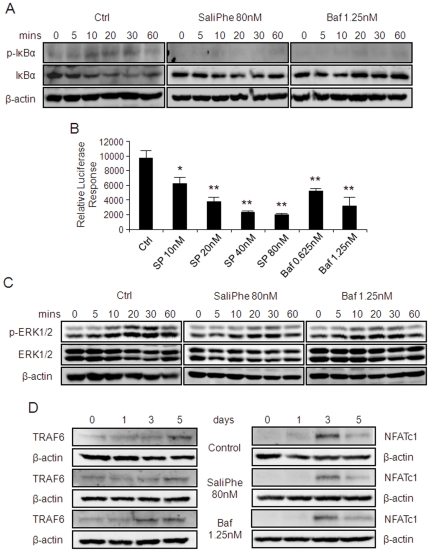
Figure 6. SaliPhe and bafilomycin impair the NF-κB and ERK1/2 signalling pathway.
(A) The effect of saliPhe and bafilomycin on RANKL-induced IκBα phosphorylation and degradation. M-CSF-dependent BMMs pretreated with or without saliPhe (80 nM) or bafilomycin (1.25 nM) were stimulated with 100 ng/ml RANKL for the indicated duration. Cells were lysed and analysed by western blot using anti-p-IκBα and anti-IκBα antibody. Antibody against β-actin was used as loading control. (B) The effect of saliPhe and bafilomycin on NF-κB activation. RAW264.7 cells stably transfected with the p-NF-κB-TA-Luc luciferase reporter gene were pretreated with saliPhe (80 nM) or bafilomycin (1.25 nM) prior to stimulation with RANKL (100 ng/ml) for 8 hrs. Cell lysates were harvested and luciferase activity was measured. Relative luciferase response was plotted. Data representative of 3 independent experiments performed in triplicates (mean ± SEM; *P<0.05, **P<0.01 against control). (C) The effect of saliPhe and bafilomycin on ERK1/2 signalling pathway. BMMs pretreated with or without saliPhe (80 nM) or bafilomycin (1.25 nM) were stimulated by RANKL for the indicated duration. The expression of ERK1/2 and phosphorylated ERK1/2 were probed using corresponding antibodies. (D) The effect of saliPhe and bafilomycin on TRAF6 and NFATc1 induction. M-CSF-dependent BMMs pretreated with or without saliPhe (80 nM) or bafilomycin A1 (1.25 nM) was stimulated with 100 ng/ml RANKL for the indicated duration. Cells were lysed and analysed by western blot using anti-TRAF6 and anti-NFATc1 antibody. Antibody against β-actin was used as loading control.

