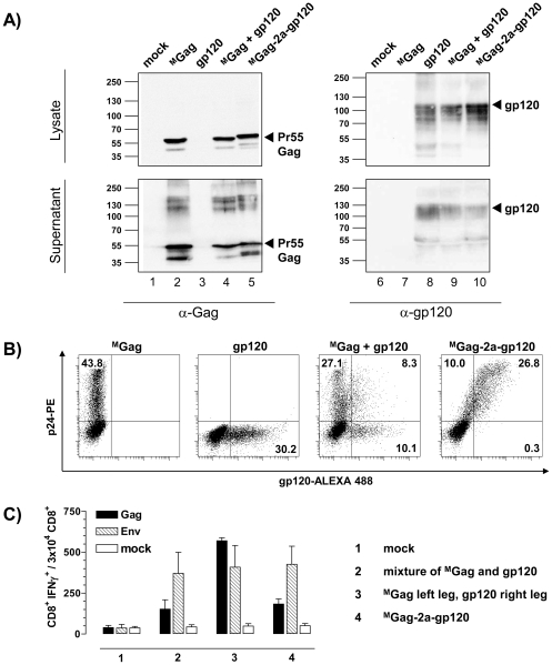
Figure 7. Expression of Gag and Env in co-transfected 293T cells and the effect of administrating Gag-2a-Env on Gag- and Env-specific CD8+ T cell responses.
(A) 293T cells were transiently transfected with 3 µg of the indicated plasmid DNA constructs. Following 48 h incubation, TCA-precipitated supernatants and cell lysates were separated by SDS-PAGE and analyzed by immunoblotting using p24-specific (Gag) mouse mAb (CB-4/1), and gp120-specific mouse mAb (MH23), as indicated. Molecular weight markers and positions of the detected proteins are indicated. mock: non-transfected 293T cells. (B) 293T cells were transiently transfected with equimolar amounts of the indicated plasmid DNA constructs. At 48 h post transfection, cells were permeabilized, stained with anti-p24-PE and/or anti-gp120-ALEXA 488 and analyzed by flow cytometry. (C) BALB/c mice (n = 6 per group) were inoculated i.m. with (i) an equimolar mixture of MGag and gp120 in both legs, (ii) MGag in the left, and gp120 in the right leg, or (iii) MGag-2a-gp120. After 12 days, spleen cells were isolated and tested for specific cellular immune responses by measuring IFNγ production after stimulation with Gag (black) and Env (hatched) specific peptides as above. IFNγ production was determined using FACS analysis after intracellular staining of IFNγ. Cell culture medium served as negative control. Data shown are representative of two experiments.

