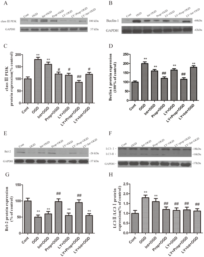
Figure 5. Blockade of the inhibition of autophagy activation by propofol.
PC12 cells were treated with propofol and the class III PI3K relatively selective inhibitor LY294002 (50 µmol/L) during the OGD insult and were harvested 6 h later for western blot analysis. (A, B, E, F) Immunoblot analyses of OGD-injured PC12 cells. The PC12 cell homogenates were analyzed by western blotting using a specific antibody against each autophagy-related protein. The expression of GAPDH was also examined for the protein loading control. (C, D, G, H) Quantification of class III PI3K, Beclin-1, Bcl-2, LC3-I and LC3-II expression. Each protein (class III PI3K,class III PI3K, Beclin-1, Bcl-2, LC3-Ι and LC3-II) shown in Fig. 5A, B, E, F was quantified after a densitometric scan and normalized to GAPDH. The optical densities of the respective protein bands were analyzed using Sigma Scan Pro 5 and normalized to the loading control (GAPDH). The results are expressed as the mean ± SD from three independent experiments. Statistical comparisons were conducted using an ANOVA followed by the Tukey test. **p < 0.01 vs. control group; # p < 0.05, ## p < 0.01 vs. OGD-treated group.

