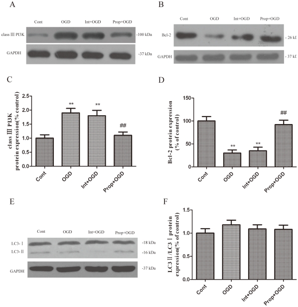
Figure 7. The expression of autophagy-related proteins during 6 h of OGD after transfection with Beclin-1 siRNA at specific concentration (100 nM) for 48 h.
(A, B, E) Immunoblot analyses of OGD-injured PC12 cells. The PC12 cell homogenates were analyzed by western blotting using a specific antibody against each autophagy-related protein. The expression of GAPDH was also examined as the protein loading control. (C, D, F) The quantification of class III PI3K, Bcl-2, LC3-I and LC3-II expression. Each protein (class III PI3K, Bcl-2, LC3-I and LC3-II) shown in Fig. 7A, B, E was quantified after a densitometric scan and normalized to GAPDH. The optical densities of the respective protein bands were analyzed using Sigma Scan Pro 5 and normalized to the loading control (GAPDH). The results are expressed as the mean ± SD from three independent experiments. Statistical comparisons were conducted using an ANOVA followed by the Tukey test. *p < 0.05, **p < 0.01 vs. the 0 hour group.

