Fig. 6.
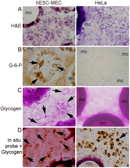
Identification of transplanted cells. (A) hESC-MECs and HeLa cells with mouse stroma adjacent to microcarriers (mc); hematoxylin staining. (B) G-6-P shown by enzyme histochemistry in transplanted hESC-MECs (arrows, brown cytoplasm). G-6-P is not expressed in HeLa cells. (C) Glycogen is present in hESC-MECs (arrows, pink cytoplasm), but is absent in HeLa cells, as expected. (D) In situ hybridization with pancentromeric human probe combined with glycogen staining to verify presence of transplanted cells. These are covered with dark hybridization signals (arrows). Magnification: ×600.

