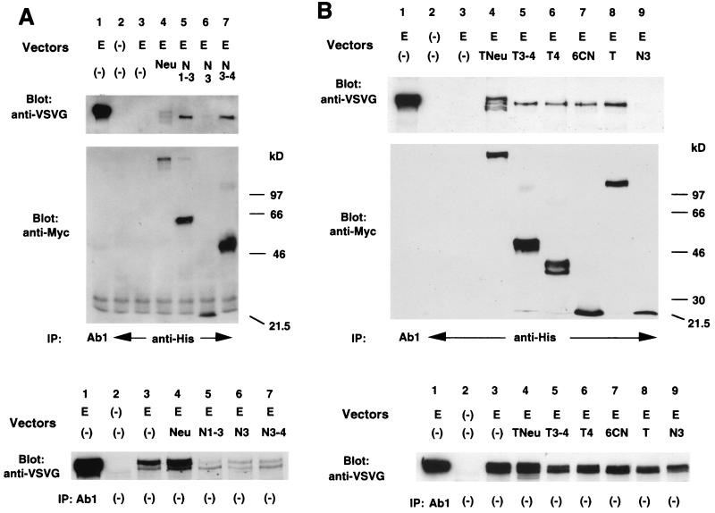
Figure 2.
Analysis of heterodimer formation between EGFR and each mutant p185c-neu protein. Cos7 cells transiently transfected with the indicated vectors were stimulated with EGF followed by the crosslinker treatment as described in Materials and Methods. Cell lysates were immunopretipitated (IP) with the antibodies as indicated. Immunoblottings (Blot) were performed with the antibodies as indicated. E, pMVEGFR; Neu, pNeu; N, pNex; N3–4, pNexD3–4; N1–3, pNex1–3; N3, pNex3; Tex, pTex; T3–4, pTexD3–4; T4, pTexD4; 6CN, pTex6CN. Membranes indicated (Top) were reblotted with anti-VSVG as indicated (Middle), and the same total cell lysates was subjected to the nitrocellulose membrane to determine the exogenous expression of EGFR (Bottom).

