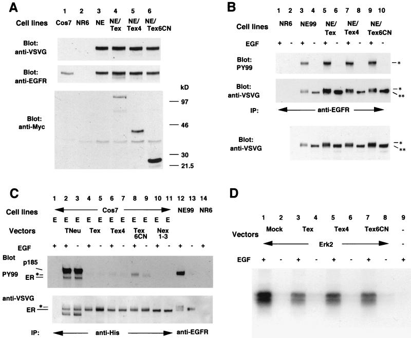
Figure 3.
Analysis of the effect of each mutant p185neu form on EGF-mediated signaling. (A) Analysis of the expression of EGFR and each mutant p185neu form in each cell line. The antibodies used were anti-VSVG (Top) followed by the second immunoblot with anti-EGFR, 1005 (Santa Cruz Biotechnology, Middle). Another membrane was immunoblotted with anti-Myc to determine the expression of each mutant p185neu form (Bottom). (B) Phosphorylation of EGFR in each NE99 clone with (+) or without (−) EGF stimulation. Membrane was immunoblotted with anti-VSVG (Middle), then reblotted with antiphosphotyrosine antibody, PY99 (Top). The same total cell lysates were transferred to the nitrocellulose membrane and blotted with anti-VSVG (Bottom). IP, immunoprecipitation; **, EGFR species with an approximate molecular mass of 170 kDa; *, the slower-migrating EGFR species. (C) Phosphorylation of heteromeric EGFR in transiently transfected Cos7 cells with (+) or without (−) EGF stimulation followed by the crosslinker treatment. Membrane was immunoblotted with anti-VSVG (Bottom), then reblotted with antiphosphotyrosine antibody, PY99 (Middle). E, pMVEGFR; ER, EGFR species with an approximate molecular mass of 170 kDa; *, slower-migrating EGFR species. (D) MAP kinase assay. Transiently transfected Cos7 cells with (+) or without (−) EGF stimulation were used for MAP kinase assay as described in Materials and Methods. Phosphorylated myelin basic protein was visualized by autoradiography. Mock, pSectagC as an empty vector; Erk2, pCDNA3-HA-ERK2.

